Atlas of PET-CT Imaging in Oncology: A Case-Based Guide to Image Interpretation
[amazon_link asins=’331918993X’ template=’ProductAd’ store=’aishabano-20′ marketplace=’US’ link_id=’6cc4f487-2049-4c91-a33f-4a9d4cceb5d0′]

Atlas of PET-CT Imaging in Oncology: A Case-Based Guide to Image Interpretation

DOWNLOAD THIS MEDICAL BOOK HERE

https://upsto.re/CKKpLqG

This atlas is a case-based guide to the interpretation of FDG PET-CT images in clinical scenarios faced by physicians during the routine practice of oncology. The book aims to help the practitioner to overcome diagnostic dilemmas through familiarization with the physiologic distribution of FDG, normal variants and benign findings. The main focus, however, is the imaging of major oncological diseases. Different pathologies are addressed in individual chapters comprising teaching files of cases, each of which corresponds to a common indication for PET-CT imaging, such as metabolic characterization of lesions, staging, restaging and evaluation of response to therapy. Each case is accompanied by an explanation of the patient’s history, interpretation of the PET-CT study, and a teaching point often supported by relevant literature. This book will be of great value to residents and practitioners in nuclear medicine, radiology, oncology, radiation oncology and nuclear medicine technology.

Clinical Atlas of Cardiac and Aortic CT and MRI 1st ed

[amazon_link asins=’3030036812′ template=’ProductAd’ store=’aishabano-20′ marketplace=’US’ link_id=’6b50b34e-69dd-4aaa-8263-6ceb9c410d62′]

Clinical Atlas of Cardiac and Aortic CT and MRI 1st ed

DOWNLOAD THIS MEDICAL BOOK HERE

https://upsto.re/CtgfJhP

This atlas comprehensively describes the application of computed tomography (CT) and magnetic resonance (MR) imaging in real-world scenarios using 192 illustrative clinical cases. These imaging techniques are revolutionizing the diagnostic and therapeutic approach for cardiovascular patients and are progressively becoming viable sub-specialties among radiologists and cardiologists.

Clinical Atlas of Cardiac and Aortic CT and MRI features clinically relevant case-based examples of how CT and MR imaging techniques can be applied to identify the pathological features of a range of acquired and congenital heart diseases. Using more than 1000 high-quality figures of distinctive CT and MR imaging features of most cardiovascular diseases, both acquired and congenital, it therefore provides a valuable resource for both specialist and non-specialist radiology/cardiology practitioners seeking to develop a deep understanding of how to recognize the features of a variety of heart diseases using CT and MR imaging techniques.

Atlas on X-ray and Angiographic Anatomy 1st Edition

[amazon_link asins=’9350904322′ template=’ProductAd’ store=’aishabano-20′ marketplace=’US’ link_id=’5933befa-a4f2-4c61-a755-fd79c3050510′]

Atlas on X-ray and Angiographic Anatomy 1st Edition

DOWNLOAD THIS MEDICAL BOOK FREE HERE

https://upsto.re/CDhxVeW

This atlas presents trainees with numerous X-ray and angiographic images to gain a thorough understanding of normal radiographic anatomy in order to make an accurate diagnosis of underlying pathology.

Presented in an easy to read format, the book covers radiological procedures, ossification centres, X-ray production, digital subtraction angiography, and computed and digital radiography, in the different anatomical sections of the body.

This practical guide includes nearly 240 clearly labelled images, illustrations and tables, with detailed descriptions, to assist learning.

Key points

  • Atlas of X-ray and angiographic images to help trainees understand normal radiographic anatomy and diagnose underlying pathology
  • Easy to read format
  • Covers different imaging techniques for all areas of the body
  • Includes nearly 240 images, illustrations and tables with detailed descriptions

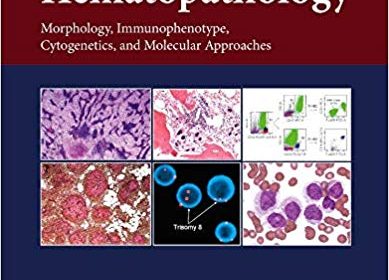
Atlas of Hematopathology: Morphology, Immunophenotype, Cytogenetics, and Molecular Approaches 2nd Edition

[amazon_link asins=’0128098430′ template=’ProductAd’ store=’aishabano-20′ marketplace=’US’ link_id=’0e4765a8-5025-452f-b570-6216833b8d75′]

Atlas of Hematopathology: Morphology, Immunophenotype, Cytogenetics, and Molecular Approaches 2nd Edition

DOWNLOAD THIS BOOK FREE HERE

https://upsto.re/CFtyBwj

Atlas of Hematopathology: Morphology, Immunophenotype, Cytogenetics, and Molecular Approaches, Second Edition, will appeal to both a wide range of people undergoing training in a variety of medical fields and practicing non-hematopathologists. For clinicians, fellows and residents, correct diagnosis (and therefore correct treatment) of diseases depends on a strong understanding of the molecular basis for the disease, making this book a crucial resource. This atlas contains hundreds of high-quality color images that mirror the findings that fellows and clinicians encounter in practice. In addition, it provides information in a quick, simple and user-friendly manner, attracting both those in training and non- experts.

Residents, fellows, practicing clinicians, and researchers in pathology, hematology and hematology/oncology will find this a useful resource.

  • Covers both non-neoplastic hematopathology and neoplastic hematopathology in all organs and tissues
  • Demonstrates results of important complementary diagnostic tests, such as immunophenotyping (immunohistochemical stains and flow cytometry), karyotyping, FISH and DNA/molecular studies
  • Provides tables to summarize the most important clinicopathological features
  • Saves clinicians and researchers time in quickly accessing the very latest details on the diverse clinical and scientific aspects of hematopathology

Atlas of Acoustic Neurinoma Microsurgery

[amazon_link asins=’3131102810′ template=’ProductAd’ store=’aishabano-20′ marketplace=’US’ link_id=’23241fa5-d3a7-4bda-9f00-6d1768889a0f’]

Atlas of Acoustic Neurinoma Microsurgery

DOWNLOAD THIS BOOK FREE HERE

https://upsto.re/DL2s9wW

This colour atlas provides information on all major acoustic neurinoma procedures, and is designed to improve physicians’ skills in this specialty. With more than 50,000 new cases per year worldwide, acoustic neurinomas are among the most commonly observed skull-base tumours. They are also among the most demanding for surgeons, since great precision is needed to safeguard the auditory nerve, the facial nerve and other critical structures. The book begins with sections on the radiographic assessment of tumours, the selection of surgical approach, and pre-operative preparations. It then turns to the main surgical approaches, using hundreds of photographs – taken during actual surgery – to lead readers through each procedure in step-by-step detail. There is a special section on the use of endoscopy in acoustic neurinoma surgery, and an analysis of 50 MR scans. This practical volume should be of interest to otolaryngologists, neurosurgeons and other physicians involved in acoustic neurinoma surgery.

Pocket Atlas of Echocardiography 1st Edition

[amazon_link asins=’3131412410′ template=’ProductAd’ store=’aishabano-20′ marketplace=’US’ link_id=’2ec1fc63-6493-4822-80f7-aa23e88ba836′]

Pocket Atlas of Echocardiography 1st Edition

DOWNLOAD THIS BOOK FREE HERE

https://upsto.re/DL2A4nL

In diagnostic cardiology, the usefulness and effectiveness of
state-of-the-art echocardiography is unsurpassed. This handy atlas includes all
of the information you need to navigate the numerous imaging planes that
transect the heart with ease and confidence.

Comprehensive coverage:


  • More than 400 illustrations, including, sharp, clear
    echocardiograms, full-color schematic diagrams, and 3-D images.
  • Detailed descriptions of all of the acoustic windows
    and imaging planes for every echocardiogram.
  • All major diseases depicted in B-mode, M-mode,
    Doppler and color Doppler.
  • A practical overview of the patient examination,
    including imaging and patient positioning.
  • All cardiac diseases are shown – valvular heart
    disease, coronary heart disease, cardiomyopathies, prosthetic valves,
    carditis, septal defects, hypertensive heart diseases, intracardiac masses.
  • Hundreds of vivid mnemonic devices and useful tips to
    help you locate, name, and remember all anatomical structures and features.

Color Atlas of Ultrasound Anatomy

[amazon_link asins=’3131390522′ template=’ProductAd’ store=’aishabano-20′ marketplace=’US’ link_id=’89812f44-fb8f-11e8-bf69-e77a2a205d65′]

Color Atlas of Ultrasound Anatomy

DOWNLOAD THIS BOOK FREE HERE

https://upstore.net/DrEWTd6

Color Atlas of Ultrasound Anatomy, Second Edition presents a systematic, step-by-step introduction to normal sectional anatomy of the abdominal and pelvic organs and thyroid gland, essential for recognizing the anatomic landmarks and variations seen on ultrasound. Its convenient, double-page format, with more than 250 image quartets showing ultrasound images on the left and explanatory drawings on the right, is ideal for rapid comprehension. In addition, each image is accompanied by a line drawing indicating the position of the transducer on the body and a 3-D diagram demonstrating the location of the scanning plane in each organ.

Special features:

More than 60 new ultrasound images in the second edition that were obtained with state-of-the-art equipment for the highest quality resolution A helpful foundation on standard sectional planes for abdominal scanning, with full-color photographs demonstrating probe placement on the body and diagrams of organs shown Front and back cover flaps displaying normal sonographic dimensions of organs for easy reference

Covering all relevant anatomic markers, measurable parameters, and normal values, and including both transverse and longitudinal scans, this pocket-sized reference is an essential learning tool for medical students, radiology residents, ultrasound technicians, and medical sonographers.

Atlas of Dermatopathology: Tumors, Nevi, and Cysts

[amazon_link asins=’1119371546′ template=’ProductAd’ store=’aishabano-20′ marketplace=’US’ link_id=’72e23242-f616-11e8-9198-8b906b479503′]

Atlas of Dermatopathology: Tumors, Nevi, and Cysts

DOWNLOAD THIS BOOK FREE HERE

https://upstore.net/Dp9GBR6

This essential guide offers trainees and practicing dermatologists alike a reliable and easy-to-navigate reference tool for the accurate and expedient diagnosis of skin diseases. With its balanced consideration of both clinical and histopathological features, the book supports the straightforward, pattern-based identification of a wide range of disorders, and breaks diagnostic criteria down into accessible, bite-sized pointers. 250 high-quality images complement the text to wonderful effect, and offer invaluable points of comparison for anyone working in the field of dermatology today.

Atlas of Hematopathology 2nd Edition

[amazon_link asins=’0128098430′ template=’ProductAd’ store=’aishabano-20′ marketplace=’US’ link_id=’eda6b2a8-ec25-11e8-8f36-cf0b2ab0979c’]

Atlas of Hematopathology: Morphology, Immunophenotype, Cytogenetics, and Molecular Approaches 2nd Edition

DOWNLOAD THIS BOOK FREE HERE

https://upsto.re/DzaSsLr

Atlas of Hematopathology: Morphology, Immunophenotype, Cytogenetics, and Molecular Approaches, Second Edition, will appeal to both a wide range of people undergoing training in a variety of medical fields and practicing non-hematopathologists. For clinicians, fellows and residents, correct diagnosis (and therefore correct treatment) of diseases depends on a strong understanding of the molecular basis for the disease, making this book a crucial resource. This atlas contains hundreds of high-quality color images that mirror the findings that fellows and clinicians encounter in practice. In addition, it provides information in a quick, simple and user-friendly manner, attracting both those in training and non- experts.

Residents, fellows, practicing clinicians, and researchers in pathology, hematology and hematology/oncology will find this a useful resource.

  • Covers both non-neoplastic hematopathology and neoplastic hematopathology in all organs and tissues
  • Demonstrates results of important complementary diagnostic tests, such as immunophenotyping (immunohistochemical stains and flow cytometry), karyotyping, FISH and DNA/molecular studies
  • Provides tables to summarize the most important clinicopathological features
  • Saves clinicians and researchers time in quickly accessing the very latest details on the diverse clinical and scientific aspects of hematopathology

Atlas of Emergency Medicine 4th Edition

[amazon_link asins=’0071797254′ template=’ProductAd’ store=’aishabano-20′ marketplace=’US’ link_id=’e7744210-de17-11e8-8f31-d36171f2b112′]

Atlas of Emergency Medicine 4th Edition

DOWNLOAD THIS BOOK FREE HERE

https://upsto.re/DTXpx5a

The most complete and trusted visual compendium of emergency medicine ― extensively updated with more than 1500 full-color illustrations.

A Doody’s Core Title for 2017!

Now in its fourth edition, The Atlas of Emergency Medicine remains the best guide to visual diagnosis of acute medical problems encountered in emergency practice. Packed with more than 1500 images, 500 new to this edition, it is the most comprehensive source of high quality emergency medicine images available, and enhanced with clear clinical information on medical emergencies.

Organized by organ-system/special populations/general issues and then by problem, The Atlas of Emergency Medicine is your “look quick, act fast” reference designed to help assess, diagnose, and treat patients efficiently. It covers basic and subtle diagnosis of a broad spectrum of typical and atypical conditions.

The book’s format is ideally suited to visual learning. One to four images per topic show you exactly what to look for. Accompanying the images is succinct “need-to-know” information for each clinical problem, including management options and clinical pearls. All treatment and diagnostic testing information and guidelines are fully updated.

NEW to this edition is the inclusion of more than 50 video clips that highlight key visual topics in emergency medicine.

1 2 3 4 5 6 19