Atlas of Acquired Cardiovascular Disease Imaging in Children 1st ed. 2017 Edition
[amazon template=iframe image2&asin=3319441132]

This atlas is a concise visual guide to the imaging of acquired heart disease in infants, children, and adolescents. Imaging plays an ever-increasing vital role in diagnosis, preoperative planning, and postoperative management for children with these disorders. The book reviews techniques for lowering radiation, discusses protocols for imaging in children, and provides recommendations for the most appropriate studies that decrease the time and cost of imaging these patients. Focusing on functional and anatomic imaging with an emphasis on three-dimensional color-coded models derived from CT and MR scans, this book promotes understanding of cardiovascular disorders in children, including infectious, neoplastic, and metabolic diseases. Atlas of Acquired Cardiovascular Disease Imaging in Children is a valuable resource through which cardiologists, radiologists, pediatric cardiothoracic surgeons, and residents can improve the quality and treatment of pediatric and adolescent patients with acquired heart disease.

DOWNLOAD THIS BOOK FREE HERE

http://upsto.re/F3xzmvU

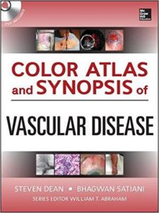
Color Atlas and Synopsis of Vascular Disease (Cardiolgy) 1st Edition

[amazon template=iframe image2&asin=0071749543]

Hundreds of High-Quality Images Span the Entire Spectrum of Vascular Disease

INCLUDES DVD WITH IMAGE BANK

Color Atlas of Vascular Diseases is designed to help you properly identify and manage vascular diseases involving the arterial, venous, and lymphatic system. It offers a clinical and synoptic approach to the full range of diagnostic and therapeutic considerations in vascular diseases. You will find hundreds of state-of-the-art clinical, diagnostic, and interventional images, along with concise, bulleted text detailing the diagnosis and patient management.

This unique combination atlas and learning text written by experts in the field, integrates the fields of vascular medicine, vascular surgery, and endovascular therapy in an effective, easy-to-learn fashion. Chapters feature succinct bulleted teaching points combined with photographs of vascular pathology. When appropriate, vivid arteriographic and ultrasonographic images are also included.

Topics include:

  • Lower Extremity Arterial Disease
  • Aortic and Upper Extremity Arterial Disease
  • Carotid Artery Occlusive Disease
  • Aneurysmal Disease
  • Non-Atherosclerotic Disorders
  • Arteriovenous Visceral Disease
  • Venous Disease
  • Limb Swelling
  • Vasospastic and Vasculitic Diseases
  • Environmental Diseases
  • Limb Ulcerations

DOWNLOAD THIS BOOK FREE HERE

http://upsto.re/FECgqMJ

Color Atlas and Synopsis of Vascular Disease (Cardiolgy) 1st Edition

[amazon template=iframe image2&asin=0071749543]

Hundreds of High-Quality Images Span the Entire Spectrum of Vascular Disease

INCLUDES DVD WITH IMAGE BANK

Color Atlas of Vascular Diseases is designed to help you properly identify and manage vascular diseases involving the arterial, venous, and lymphatic system. It offers a clinical and synoptic approach to the full range of diagnostic and therapeutic considerations in vascular diseases. You will find hundreds of state-of-the-art clinical, diagnostic, and interventional images, along with concise, bulleted text detailing the diagnosis and patient management.

This unique combination atlas and learning text written by experts in the field, integrates the fields of vascular medicine, vascular surgery, and endovascular therapy in an effective, easy-to-learn fashion. Chapters feature succinct bulleted teaching points combined with photographs of vascular pathology. When appropriate, vivid arteriographic and ultrasonographic images are also included.

Topics include:

  • Lower Extremity Arterial Disease
  • Aortic and Upper Extremity Arterial Disease
  • Carotid Artery Occlusive Disease
  • Aneurysmal Disease
  • Non-Atherosclerotic Disorders
  • Arteriovenous Visceral Disease
  • Venous Disease
  • Limb Swelling
  • Vasospastic and Vasculitic Diseases
  • Environmental Diseases
  • Limb Ulcerations

DOWNLOAD THIS BOOK FREE HERE

http://upsto.re/FQgmZG3

Normal and Abnormal Fetal Face Atlas: Ultrasonographic Features 1st ed. 2017 Edition

[amazon template=iframe image2&asin=3319437682]

This atlas offers a guide to studying the fetal face by means of ultrasound analysis. It describes key phases in cranial-facial development, allowing the reader to learn the related semiology from its most simple iteration to the most complex one.
The overall examination of a newborn’s face offers a rich source of information and can guide the general examination. The same applies in the context of fetal ultrasound examination. The analytical study of the fetal face not only makes it possible to screen for anomalies related to the face itself, but also yields valuable insights into the brain, the limbs, and the heart. In addition, it allows ultra-sonographers to unravel the puzzle of fetal dimorphism.
Written in a pedagogical style, the book guides walks the reader through the diagnostic reasoning process step by step.
The authors are pioneers in this field and teach in various university and master’s degree ultrasound programs. Their aim is to share with readers their diagnostic approaches and their knowledge and passion for 2D and 3D ultrasound techniques. Each chapter includes algorithms, biometry curves, and simple guidelines that allow users to go “from sign to syndrome”. The first chapter, which focuses on innovative embryology adapted to the needs of ultra-sonographers, was written by Gérard Couly, a maxilla-facial surgeon and the founding father of the specialty>

DOWNLOAD THIS BOOK FREE HERE

http://upsto.re/AoZYZPn

Atlas of Imaging in Infertility: A Complete Guide Based in Key Images 1st ed. 2017 Edition

[amazon template=iframe image2&asin=3319138928]

This atlas gathers the most frequent imaging findings concerning alterations that cause infertility in both males and in females. Also, it discusses how the images should be analyzed and described, to serve as a guide for the physicians who request and perform these imaging tests.
Infertility is a highly prevalent condition – about 10% of couples are infertile with males and females equally affected. This book offers professionals dealing with infertility, including endocrinologists, gynecologists and urologists, a useful guide to the diagnostic imaging tests that are essential in treating these patients. The Atlas of Imaging in Infertility is a valuable resource for physicians from different specialties managing cases of infertility.

DOWNLOAD THIS BOOK FREE HERE

http://upsto.re/AoUXE8y

Atlas of Craniocervical Junction and Cervical Spine Surgery 1st ed. 2017 Edition

[amazon template=iframe image2&asin=3319427350]

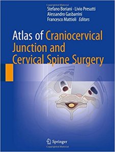
This atlas documents current surgical approaches to the craniocervical junction and the cervical spine, providing step-by-step guidance on procedures and cervical spine stabilization techniques. Opening chapters present essential information on anatomy, depict pathologies with the aid of illustrative cases, describe the role of imaging techniques in patient evaluation, and discuss surgical instrumentation and patient positioning. The different techniques employed in this delicate anatomic region, including transnasal and transoral endoscopic approaches to the craniocervical junction and posterior and anterior approaches to the cervical spine, are then explained and illustrated with a view to providing the surgeon with a clear reference that can be used in the operating room. In addition, practical advice is offered on the treatment of potential complications, postoperative management, and rehabilitation. This book will be of value not only to neurosurgeons but also to orthopedists, ENT surgeons, neurologists, and physiatrists.

DOWNLOAD THIS BOOK FREE HERE

http://upsto.re/A4mqzyz

Atlas of Differential Diagnosis in Breast Pathology (Atlas of Anatomic Pathology) 1st ed. 2017 Edition

[amazon template=iframe image2&asin=1493966952]

This atlas illustrates the range of breast lesions with detailed correlation of gross and microscopic features. Where relevant, radiological images are incorporated.  A description of normal, developmental and physiological breast morphology will serve as introduction to the main content of this atlas. Classification of tumors is based on the latest World Health Organization Classification of Tumors of the Breast, 4th edition, 2012. As immunohistochemistry is a key adjunctive tool in the workup of breast lesions as well as used in prognostic evaluation of breast cancers, appropriate examples are interspersed among the lesions where pertinent.

DOWNLOAD THIS BOOK FREE HERE

http://upsto.re/Ay6uvnz

Atlas of Clinical Diagnosis, 2e 2nd Edition

[amazon template=iframe image2&asin=0702026689]

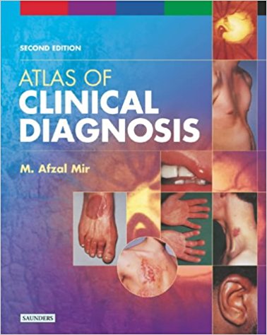
The new, 2nd Edition of this practical guide employs a sign-oriented approach to the diagnosis of both common and rare medical disorders. This comprehensive collection of clinical pictures, organized by anatomical region, reflects actual clinical practice where a diagnosis has to be constructed from a single sign or a collection of signs. Fundamental, pathognomonic, and commonly seen specific and non-specific clinical signs are presented with clinical features highlighted for rapid recognition and interpretation. In addition, the book provides guidance on appropriate bedside clinical tests for establishing diagnoses.

  • Presents over 1,300 high quality photographs―complemented by concise explanatory text―to aid in diagnosis.
  • Covers all general medical problems as well as disorders in dermatology, neurology, rheumatology, ophthalmology, and endocrinology.
  • Organized logically by anatomic area.
  • Now includes explanatory captions for all illustrations.
  • Includes a wealth of new photographs for even greater coverage of clinical conditions.
  • Features a revised and improved text design with larger pages.

DOWNLOAD THIS BOOK FREE HERE

http://upsto.re/ArMAKuD

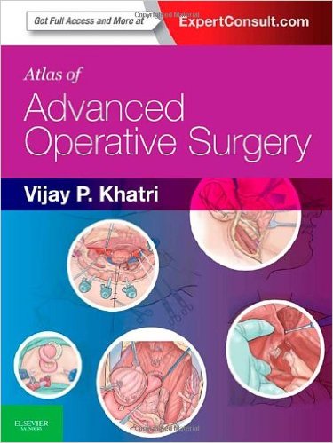
Atlas of Advanced Operative Surgery: Expert Consult – Online and Print, 1e 1 Har/Psc Edition

[amazon template=iframe image2&asin=1416041095]

Advance your surgical expertise with Atlas of Advanced Operative Surgery! This new resource picks up where other surgical references leave off, providing highly visual, step-by-step guidance on more than 100 advanced and complex procedures in both general and subspecialty areas.


  • Visualize every procedure thanks to more than 1,000 illustrations, most in full color – including intraoperative photos, beautifully illustrated color drawings that highlight the relevant anatomy and techniques in specific surgeries, and radiologic images that help you identify variations in anatomy prior to surgery.
  • Grasp each procedure and review key steps quickly with a consistent, highly focused, bulleted format.
  • See the advantages and disadvantages of variations in technique with a Pro/Con section written by expert surgeons.
  • Focus on the advanced practice skills that are of particular value to those poised to begin practice, as well as surgeons who are already in practice.
  • Benefit from the masterful guidance of Dr. Vijay Khatri, a respected expert and experienced mentor of trainees, junior faculty, and community surgeons.
  • Learn new procedures or refresh your memory on operative details prior to surgery with an easy-to-follow, step-by-step format: pre-operative preparation, operative technique, position, incision, main dissection, closure, alternative technical approaches (with pros/cons), and post-operative care.

DOWNLOAD THIS BOOK FREE HERE

http://upsto.re/Ar7RLyX

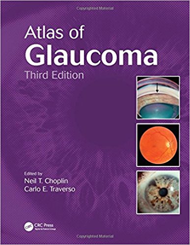
Atlas of Glaucoma, Third Edition 3rd Edition

[amazon template=iframe image2&asin=1482209217]

Glaucoma affects all age groups and is a leading cause of blindness worldwide. It is imperative that practicing clinicians and surgeons recognize both primary and secondary glaucoma as well as cases of glaucoma associated with other disorders. Atlas of Glaucoma, Third Edition provides an in-depth review and analysis of the management of glaucoma and discusses research advancements that have resulted in the latest therapies.

See What’s New in the Third Edition:

  • The influence of the cornea on intraocular pressure measurements and new devices designed to account for this
  • Spectral domain optical coherence tomography (OCT)
  • New statistical methods for tracking rates of progression
  • New medications released since 2006
  • New chapter on trabecular bypass surgery

Additional topics include:

  • Classification of glaucoma
  • Measurement of intraocular pressure and gonioscopy
  • The optic nerve
  • Psychophysical and electrophysiological testing in glaucoma
  • Primary open-angle, secondary open-angle, and angle closure glaucomas
  • Ocular blood flow and metabolism
  • Medical therapy and surgical treatments
  • Aqueous shunts
  • Combined cataract and glaucoma surgery
  • Treatment of developmental glaucoma

This colorful atlas contains detailed captions for each illustration, making it ideal for teaching purposes as well as a reference for the practicing ophthalmologist.

Glaucoma encompasses a wide variety of clinical entities, clinical findings, diagnostic techniques, and treatment options. This book presents a vast assemblage of insight from internationally recognized experts.

DOWNLOAD THIS BOOK FREE HERE

http://upsto.re/ArfiXkb

1 14 15 16 17 18 19