Fundamentals of Sectional Anatomy: An Imaging Approach 2nd Edition

[amazon_link asins=’1133960863′ template=’ProductAd’ store=’aishabano-20′ marketplace=’US’ link_id=’64264095-e1ec-11e8-8483-61afb5fae201′]

Fundamentals of Sectional Anatomy: An Imaging Approach 2nd Edition

DOWNLOAD THIS BOOK FREE HERE

https://upsto.re/DmYPpVf

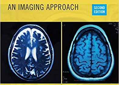
The second edition of Fundamentals of Sectional Anatomy: An Imaging Approach is the ideal introductory text for new radiography students, seasoned students preparing for the CT and MRI exams, or anyone interested in learning about human anatomy. Chapters address the fundamentals of sectional anatomy, starting at the vertex of the skull and descending to the symphysis pubis, with additional in-depth coverage of the vertical column, major joints of the upper and lower extremities, and separate chapters on the facial bones and sinuses. This systematic approach to the organization of the book provides students with the most complete presentation and realistic exposure to sectional anatomy available. Numerous line drawings and two complete sets of fully labeled images complement each section of the text to strengthen the learning experience, while end-of-chapter summaries and review questions challenge readers to assess their understanding of important topics. Building upon its reputation for an uncluttered presentation and clearly labeled images, this new edition presents more than 200 new MR images, dozens of CT images, and new complex illustrations–transporting this already fascinating book into the modern age of radiography.

USMLE Step 1 Lecture Notes 2018: Anatomy (USMLE Prep)

[amazon_link asins=’1506220924′ template=’ProductAd’ store=’aishabano-20′ marketplace=’US’ link_id=’19f4398d-e1eb-11e8-ac4f-8b27f0e2adcd’]

USMLE Step 1 Lecture Notes 2018: Anatomy (USMLE Prep)

DOWNLOAD THIS BOOK FREE HERE

https://upsto.re/DmaToJp

Kaplan Medical’s USMLE Step 1 Lecture Notes 2018: Anatomy offers in-depth review with a focus on high-yield topics – a comprehensive approach that will help you deepen your understanding while focusing your efforts where they’ll count the most.

Used by thousands of medical students each year to succeed on USMLE Step 1, Kaplan’s official lecture notes are packed with full-color diagrams and clear review.

The Best Review

  • Organized in outline format with high-yield summary boxes for efficient study.
  • Clinical correlations and bridges between disciplines highlighted throughout.
  • Full-color diagrams and charts for better comprehension and retention.
  • Updated annually by Kaplan’s all-star expert faculty

Grant’s Atlas of Anatomy (Grant, John Charles Boileau//Grant’s Atlas of Anatomy) Fourteenth, North American Edition

[amazon_link asins=’1469890682′ template=’ProductAd’ store=’aishabano-20′ marketplace=’US’ link_id=’c41af221-dc73-11e8-9636-1ff7e7f02563′]

Grant’s Atlas of Anatomy (Grant, John Charles Boileau//Grant’s Atlas of Anatomy) Fourteenth, North American Edition

DOWNLOAD THIS BOOK FREE HERE

https://upsto.re/DJnzYGu

Illustrations drawn from real specimens, presented in surface-to-deep dissection sequence, set Grant’s Atlas of Anatomy apart as the most accurate reference available for learning human anatomy. These realistic representations provide students with the ultimate lab resource.

For more than seventy years, Grant’s Atlas of Anatomy has continually adapted to meet the needs of each generation of students, while maintaining the Grant’s tradition of excellence. The meticulously updated fourteenth edition is a visually stunning reference that delivers the accuracy, pedagogy, and clinical relevance expected of this classic atlas, with new features that make it even more practical and user-friendly:

  • Recolorization of the original Grant’s Atlas images from high-resolution scans add a new level of organ luminosity and tissue transparency, and enable the formation of 3D constructs for each body region. This lavish visual enhancement ensures a vastly improved experience in learning the structure and function of the human body. See the enhanced color and detail here:  https://youtu.be/KdLzQUoG54Y
  • Schematic illustrations now feature a uniform style and consistent coloring, clearly showing the relationships of structures and clarifying anatomical concepts.
  • Newly revised legends with highlighted clinical applications describe the anatomic features and provide context for health care practice.
  • Enhanced medical imaging includes more than 100 clinically significant MRIs, CT images, ultrasound scans, and corresponding orientation drawings.
  • Updated and improved tables help organize muscles, vessels, and other anatomic information in an easy-to-use format ideal for review and study.

Color Atlas of Human Anatomy: Vol. 2: Internal Organs 6th edition Edition

[amazon_link asins=’3135334066′ template=’ProductAd’ store=’aishabano-20′ marketplace=’US’ link_id=’ac657859-bf9a-11e8-b10d-27090ffc586e’]

Color Atlas of Human Anatomy: Vol. 2: Internal Organs 6th edition Edition

DOWNLOAD THIS BOOK FREE HERE

https://upsto.re/DQFy72u

The sixth edition of this classic work makes mastering a vast amount of information on internal organs much less daunting. It offers a vivid review of the human body and its structure, and it is an ideal study companion as well as an excellent basic reference text.

These are some of the many user-friendly features of this book

  • New color plates on embryology and histology
  • More than 200 outstanding full-color illustrations and 130 clinical correlations
  • Side-by-side images with explanatory text
  • An overview of anatomical terms in each section

Emphasizing clinical anatomy, this text integrates current information from a wide range of medical disciplines into discussions of the internal organs, including:

  • Cross-sectional anatomy as a basis for working with modern imaging modalities
  • Detailed explanations of organ topography and function
  • Physiological and biochemical information included where appropriate
  • An entire chapter devoted to pregnancy and human development

Volume 2: Internal Organs and its companions Volume 1: Locomotor System and Volume 3: Nervous System and Sensory Organs comprise a must-have resource for students of medicine, dentistry, and all allied health fields.

Surgical Anatomy of the Abdomen

[amazon_link asins=’9732726148′ template=’ProductAd’ store=’aishabano-20′ marketplace=’US’ link_id=’a3d14397-bd94-11e8-bca7-5dbd87aace84′]

Surgical Anatomy of the Abdomen

DOWNLOAD THIS BOOK FREE HERE

https://upsto.re/DQnTrue

It is our privilege to introduce a book of surgical anatomy, especially during this time, when classical anatomy with thorough anatomical dissection on cadaveric models has been reduced significantly in medical schools, while the surgeon’s needs for anatomical knowledge has continuously increased. In the repertoire of modern general surgery, are included nowadays, trauma and emergency surgery, coloproctology, esophagus and cardia and stomach surgery, surgical oncology, hepato-pancreato-biliary surgery, endocrine surgery, transplant surgery and breast surgery. During the evolution of this speciality, surgical anatomy has experienced new refinements. Throughout this book we intended to fill the gap between all we are learning as students in the pre-clinical years and what we are facing everyday in the operating theaters. We feel that a major strength of this book is integration of embryology details, relevant for the operating surgeon who should develop dissection planes with minimum trauma and blood loss, according to the modern open and minimally invasive surgical principles. The histology data tries to offer to residents and young doctors a tool for better understanding the tissue structures relevant in surgical oncology. Being the first edition of this book, we are aware about the need for further clarification in certain areas and we rely on constructive criticism from our readers. The book is intended for a wide readership across students, residents, and general surgery physicians. We are grateful to our colleagues from the Emergency Hospital of Bucharest, from Surgery, Radiology and Imagistics, Pathology and Gastroenterology Departments who supported us with useful images. Many authors have given us permission to reproduce their figures and detailed acknowledgements are given in the text. The publisher, the Publishing House of the Romanian Academy, enthusiastically supported us with editing of our book and provided us with outstanding assistance. We hope that this book will not linger on the shelf and will be a constant place to return to for revising anatomy before going into the operating room.

Running Anatomy

[amazon_link asins=’1492548294′ template=’ProductAd’ store=’aishabano-20′ marketplace=’US’ link_id=’5b619077-bc7b-11e8-892a-37c480bd2fd8′]

Running Anatomy

DOWNLOAD THIS BOOK FREE HERE

https://upsto.re/DEpRb4f

Running Anatomy features 48 of the most effective strength exercises for runners, each with clear step-by-step descriptions and full-color anatomical illustrations highlighting the muscles in action. Each illustration helps you better understand how muscles, ligaments, and tendons all work together as your body moves.

Running Anatomy clearly links each exercise to running performance. You’ll see how to strengthen specific muscles and improve gait efficiency for faster times and more fluid runs. Plus you’ll learn how to eliminate anatomical imbalances that can lead to the most common injuries that runners face, including plantar fasciitis, lower-back pain, knee aches and strains, and torn muscles and tendons.

Feel prepared for any challenge that comes your way. You’ll find variations to train for every condition and competition—for various terrains, speeds, elevations, and distances, from sprint to marathon. You’ll also learn how new gear and technology-enhanced equipment can maximize your training and performance.

Whether you’re a fitness runner looking to conquer hills with more speed and strength or a competitive runner looking for that extra edge in performance and a finishing kick, Running Anatomy will ensure that you’re ready to achieve your personal best.

CE exam available! For certified professionals, a companion continuing education exam can be completed after reading this book. The Running Anatomy, Second Edition, Online CE Exammay be purchased separately or as part of the Running Anatomy, Second Edition With CE Exam, package that includes both the book and the exam.

Applied Anatomy and Physiology for Speech-Language Pathology and Audiology 1st Edition

[amazon_link asins=’0781788374′ template=’ProductAd’ store=’aishabano-20′ marketplace=’US’ link_id=’fecc3037-b793-11e8-a286-cb410ad80014′]

Applied Anatomy and Physiology for Speech-Language Pathology and Audiology 1st Edition

DOWNLOAD THIS BOOK FREE HERE

https://upsto.re/3WfiDGj

Publisher’s Note: Products purchased from 3rd Party sellers are not guaranteed by the Publisher for quality, authenticity, or access to any online entitlements included with the product.

This textbook aligns the basic science of anatomy and physiology with the applied art and science of communication disorders. The content is approached from a clinical perspective so that students understand the application of the content. Applied Anatomy and Physiology for Speech-Language Pathology and Audiology is unique for its presentation of elementary and introductory anatomy and physiology in a framework of clinical practice.

Rohen’s Photographic Anatomy Flash Cards Second Edition

[amazon_link asins=’1451194501′ template=’ProductAd’ store=’aishabano-20′ marketplace=’US’ link_id=’4ff5eaab-b790-11e8-92b2-df2b96e861e9′]

Rohen’s Photographic Anatomy Flash Cards Second Edition

DOWNLOAD THIS BOOK FREE HERE

https://upsto.re/3W5WnHZ

Derived from the leading dissection atlas Anatomy: A Photographic Atlas by Johannes W. Rohen, Chihiro Yokochi, and Elke Lütjen-Drecoll, these flash cards showcase full-color photographs of actual cadaver dissections. 220 cards, labeled for self-testing, serve as a practical tool to help students prepare for the dissection lab and the operating room.

  • Now includes review questions and additional clinical anatomy.
  • 220 full-color photographs present exquisitely dissected cadavers.
  • Ideal study aid to review recently dissected regions and to prepare for practical exams.
  • Includes a key ring and hole-punched corners for easy organization and on-the-go portability.
1 8 9 10 11 12 21