Surgical Retina (Retina Atlas)
Surgical Retina (Retina Atlas)
Surgical Retina (Retina Atlas)
DOWNLAOD THIS BOOK
Medical Books Library for Doctors, Physicians, Surgeons, Dentists, Intensivists, Physician Assistants, Nurses, Medical Technicians and Medical Students
Medical books library


Surgical Retina (Retina Atlas)
Geriatric Ophthalmology: A Competency-based Approach 2nd Edition
Geriatric patients have unique responses to treatment and disease, often harboring co-morbidities that can impact evaluation, treatment, and prognosis, which can require specialized expertise or experience. Geriatric Ophthalmology, Second Edition draws upon the successful first edition by applying a competency-based approach to these patients, improving awareness, increasing understanding, and encouraging expertise about geriatric issues among eye care professionals. These intersecting conditions and their treatment are comprehensively discussed in this fully updated second edition, complete with additional high-quality illustrations and photos. Each chapter utilizes illustrative cases to exemplify the points of care encompassed by the competencies. Topics of special interest are included, such as diabetic retinopathy, age-related macular degeneration, cataracts, glaucoma, diabetic retinopathy, low vision, all diseases of aging, and the effect of vision loss on the geriatric patient’s quality of life. Medical students, residents, fellows, clinicians, and allied health personnel alike will find this to be a comprehensive resource and exceptional guide to the care of older patients with geriatric ophthalmology problems.
Atlas of Pediatric Cataract
This book highlights various pediatric cataracts and their clinical presentations, along with a wealth of high-quality images collected over two decades. Offering a precise guide to illustrative interventions, it contains images collected pre-operatively, intra-operatively and post-operatively using slit lamp or microscope integrated cameras. The book covers congenital, developmental and traumatic cataracts, along with other rare morphologies. It presents selected challenging clinical situations, along with surgical management steps that draw on high-quality images.
Separate chapters address recent advances in the field of pediatric cataracts, e.g. ultrasound biomicroscopy, lenticular abnormalities, intraoperative rhexis assistant, plasma blade/ diathermy, vascular cautery in cases of persistent fetal vasculature, operative room aberrometry, and optiwave refractive analysis. Given its scope, the book offers a valuable resource for practicing general and pediatric ophthalmologists, researchers and students alike.
Glaucoma Drainage Devices: A Practical Illustrated Guide
This book offers a comprehensive guide to the use of glaucoma drainage devices (GDDs) in various clinical settings, and covers aspects ranging from the basics to managing complications. The aim of this work is to provide readers with a practical go-to desktop book to assist in and enhance their surgical competence with glaucoma drainage devices.
Starting with the history of GDDs, it addresses various devices, their models and modifications, and highlights their advantages and disadvantages through numerous illustrations. The indications for the drainage devices are discussed in detail, using patient cases with photographs. The book describes the techniques for all devices in detail, which are explained further in accompanyin videos.
After covering the basic techniques, the book provides extensive notes on modifications that may be required in various case presentations such as congenital glaucoma, post-penetrating keratoplasty with extensive peripheral synechiae, and procedure through pars plans etc. Complications and their management are subsequently addressed.
The book is an essential guide to help surgeons match patients to the most suitable device, and to support patients from preparation through post-operative care. Primarily intended for glaucoma surgeons, it offers a valuable resource for fellows in training, and all who have an interest in glaucoma surgery.
This second volume in a short series on the glaucomas is devoted to the two main types of the disease, open angle glaucoma and angle closure glaucoma. The contents are based on observations and experience gained during the management of more than 40,000 hospital and 7,000 private patients, with follow-up of 10 to 50 years. The book is comprehensive in scope. All aspects of diagnosis are carefully examined, surgical indications and techniques are explained, and the role of medical treatment is evaluated. Importantly, chapters are included on topics that are frequently overlooked in other books but are important to early diagnosis, such as diurnal pressure curve, HRT, and non-conventional perimetry. This volume provides a wealth of information and includes almost 1000 illustrations, the majority in color, as well as an accompanying DVD of surgical techniques. It will be an invaluable asset for all ophthalmologists.
Major Revision This year Section 6 is updated to include 37 videos, plus several new animations and images. New animations demonstrate the Krimsky test, cover-uncover test, alternate cover test, prism alternate cover test, simultaneous prism and cover test. Tables detailing neuro-oculocutaneous syndromes are also included. This volume begins with discussions of the pediatric eye examination and extraocular muscle anatomy. Includes chapters on amblyopia, sensory physiology and pathology, and the diagnostic evaluation of strabismus. Examines the clinical features, diagnosis and treatment of esodeviations and exodeviations, pattern strabismus, vertical deviations, nystagmus, and special forms of strabismus. Also, describes key aspects of extraocular muscle surgery. The second part of this book covers the full range of pediatric ocular disorders. Videos covering diagnostic evaluation, procedures for vertical deviations and surgery of the extraocular muscles are included. Both print and eBook users have access to the videos. Upon completion of Section 6, readers should be able to: *Describe evaluation techniques for young children that provide the maximum gain of information with the least trauma and frustration *Explain the classification and diagnosis of amblyopia, as well as the treatment options *Describe the commonly used diagnostic and measurement tests for strabismus *List the possible complications of strabismus surgery, and describe guidelines to minimize them *Design an approach to the diagnosis of decreased vision in children, and list resources available to these patients Major revision: 2018 2019 Section chair: Robert W. Hered, MD CME Information The American Academy of Ophthalmology is accredited by the Accreditation Council for Continuing Medical Education (ACCME) to provide continuing medical education for physicians. The American Academy of Ophthalmology designates this enduring material for a maximum of 10 AMA PRA Category 1 CreditsTM. Physicians should claim only the credit commensurate with the extent of their participation in the activity. The American Medical Association requires that all learners participating in activities involving enduring materials complete a formal assessment before claiming CME credit. To assess your achievement in this activity and ensure that a specified level of knowledge has been reached, a post-test for this section of the Basic and Clinical Science Course is provided. A minimum score of 80% must be obtained to pass the test and claim CME credit. Visit CME Central for more information. About the BCSC The Academy’s Basic and Clinical Science CourseTM (BCSC®) is ophthalmology s definitive compilation of scientific research and clinical experience. It is continually updated by a faculty of more than 100 expert ophthalmologists. Each of the 13 volumes includes fundamental clinical knowledge; numerous tables, photos and illustrations; self-assessment questions with answers; and opportunities for earning AMA PRA Category 1 CreditTM. Beginning with the 2013-2014 edition, the Academy and the European Board of Ophthalmology (EBO) have partnered to make the BCSC the standard text for all European ophthalmology training programs. The EBO now recommends the BCSC as the primary educational resource for European trainees and ophthalmologists studying for the annual EBO Diploma Exam.
Provides the essential scientific grounding for current ophthalmic practice. Discusses ocular anatomy, embryology, biochemistry and metabolism in the eye, and ocular pharmacology. Features genetics information, including an overview of appropriate uses for the latest genetic techniques. Upon completion of Section 2, readers should be able to: *Identify the bones making up the orbital walls and the orbital foramina *Identify the biochemical composition of the various parts of the eye and the eye s secretions *Demonstrate how appropriate diagnosis and management of genetic diseases can lead to better patient care *Describe the features of the eye that facilitate or impede drug delivery *Describe the mechanisms of action of antibiotic, antiviral, and antifungal medications *Discuss the anesthetic agents used in ophthalmology Last major revision: 2014-2015 Section chair: Lawrence M. Levine, MD CME Information The American Academy of Ophthalmology is accredited by the Accreditation Council for Continuing Medical Education (ACCME) to provide continuing medical education for physicians. The American Academy of Ophthalmology designates this enduring material for a maximum of 10 AMA PRA Category 1 CreditsTM. Physicians should claim only the credit commensurate with the extent of their participation in the activity. The American Medical Association requires that all learners participating in activities involving enduring materials complete a formal assessment before claiming CME credit. To assess your achievement in this activity and ensure that a specified level of knowledge has been reached, a post-test for this section of the Basic and Clinical Science Course is provided. A minimum score of 80% must be obtained to pass the test and claim CME credit. Visit CME Central for more information. About the BCSC The Academy’s Basic and Clinical Science CourseTM (BCSC®) is ophthalmology s definitive compilation of scientific research and clinical experience. It is continually updated by a faculty of more than 100 expert ophthalmologists. Each of the 13 volumes includes fundamental clinical knowledge; numerous tables, photos and illustrations; self-assessment questions with answers; and opportunities for earning AMA PRA Category 1 CreditTM. Beginning with the 2013-2014 edition, the Academy and the European Board of Ophthalmology (EBO) have partnered to make the BCSC the standard text for all European ophthalmology training programs. The EBO now recommends the BCSC as the primary educational resource for European trainees and ophthalmologists studying for the annual EBO Diploma Exam.
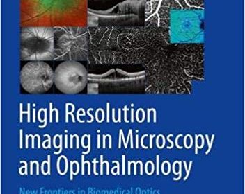
This open access book provides a comprehensive overview of the application of the newest laser and microscope/ophthalmoscope technology in the field of high resolution imaging in microscopy and ophthalmology. Starting by describing High-Resolution 3D Light Microscopy with STED and RESOLFT, the book goes on to cover retinal and anterior segment imaging and image-guided treatment and also discusses the development of adaptive optics in vision science and ophthalmology. Using an interdisciplinary approach, the reader will learn about the latest developments and most up to date technology in the field and how these translate to a medical setting.
High Resolution Imaging in Microscopy and Ophthalmology – New Frontiers in Biomedical Optics has been written by leading experts in the field and offers insights on engineering, biology, and medicine, thus being a valuable addition for scientists, engineers, and clinicians with technical and medical interest who would like to understand the equipment, the applications and the medical/biological background.
This book discusses more than one hundred patients in which visual pathway is involved, and focuses on the role of visual field examination in the diagnosis of these diseases. It also highlights the application of concepts from the new interdiscipline, integration medicine as well as molecular biology and genetics in the analysis of the diseases.
In this book, the commonly (typically) noticed changes in the visual field of patients with visual pathway disorders are mainly described in the chapter one titled as “Visual Field-related Anatomy of Visual Pathway” and chapter two titled as “Interpretation of Visual Field Test”, while the majority of the cases presented with ‘atypical’ changes in visual field. At this point, the changes in the visual field could function as either a key to understand the disease, or a question mark which confuses the diagnosis. However, the process of pushing aside a fog around the diagnosis step by step helps the readers to gradually disclose the essence of the disease.